

Modeling Disease: Wax Moulages in Early Twentieth-Century Montreal
Wax moulages—realistic wax models of pathological anatomy—became vital teaching aids in Montreal’s major teaching hospitals. Moulages have a centuries-long history of supplementing surgical and clinical teaching. In early twentieth century Montreal, these were often created by medical illustrators.
Moulages allow for close study of subtle details of disease unavailable in specimens or photographs. Typically, the creation process involved medical illustrators observing or working directly with a specimen, then creating wax models with realistic qualities that even a preserved specimen could not retain.
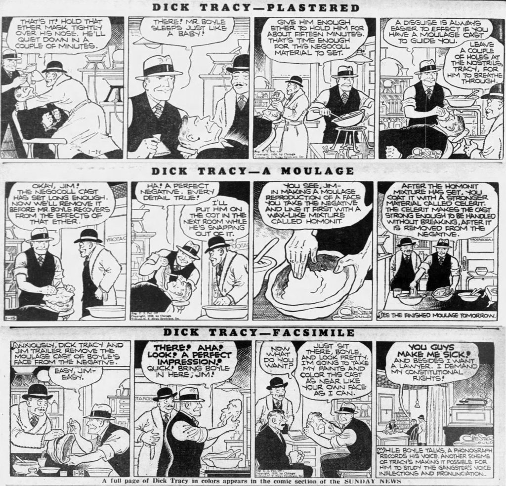
A Dick Tracy cartoon shows the process of making a moulage, a technique that fascinated both medical and popular audiences. Daily News, 14-16 January 1936.
At the Montreal General Hospital, Hortense Douglas was a key figure in this specialized craft. She not only created wax moulages, but also authored an article published by the International Association of Medical Museums, evidencing her expertise.
Douglas detailed the process of creating plaster moulds from pathological specimens for wax casting and included the field standard recipe: 2 lbs. white beeswax, 1 ¾ lbs. paraffin wax, 1 lb. talcum powder, 1 lb. prepared cornstarch, 2 oz. yellow beeswax. Pigments would be painted into the final layers before a finishing coat, and models were usually mounted to a wooden board. According to Douglas, this process allowed for endless possibilities.
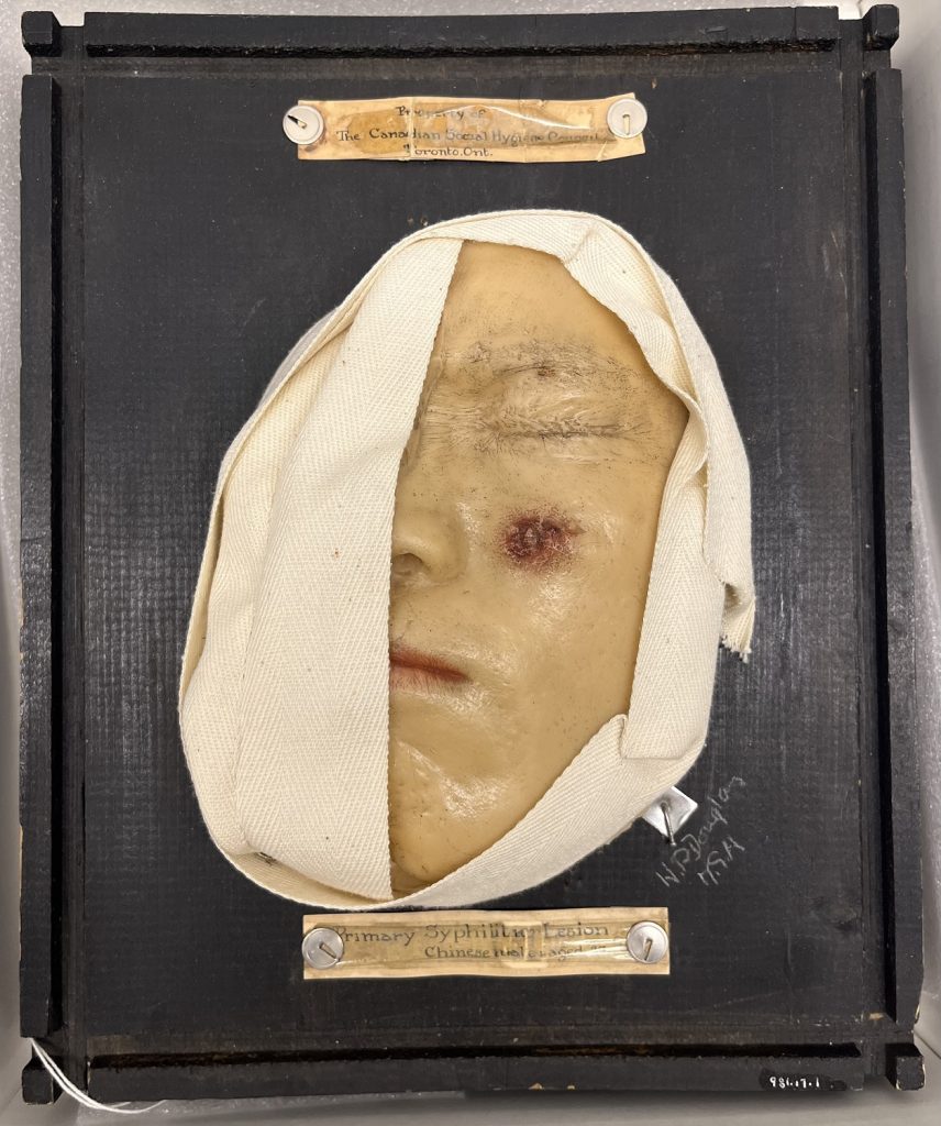
Wax moulage of syphilitic lesions created by Hortense Douglas at the Montreal General Hospital.
Harriett Blackstock worked in wax moulages during the same period in McGill’s Department of Art as Applied to Medicine. Her work was included in a 1926 exhibition for medical professionals in the McGill Medical building, where a journalist noted, “The new method of making wax models is expected to be of great value in the study of contagious diseases in McGill Medical School.”
With these two case studies, we see the rigour and precision that both medical illustrators bring to their multidisciplinary practices. The record of their work in medical journals and newspapers also underscores the value of moulages as both scientific record and pedagogical tool.
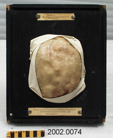
Wax moulage of syphilitic scarring created by Hortense Douglas at the Montreal General Hospital.