

MacArthur Front and Centre: Illustrating Surgery at the Royal Victoria Hospital
Helen MacArthur’s career unfolded during a pivotal shift in medical publishing.
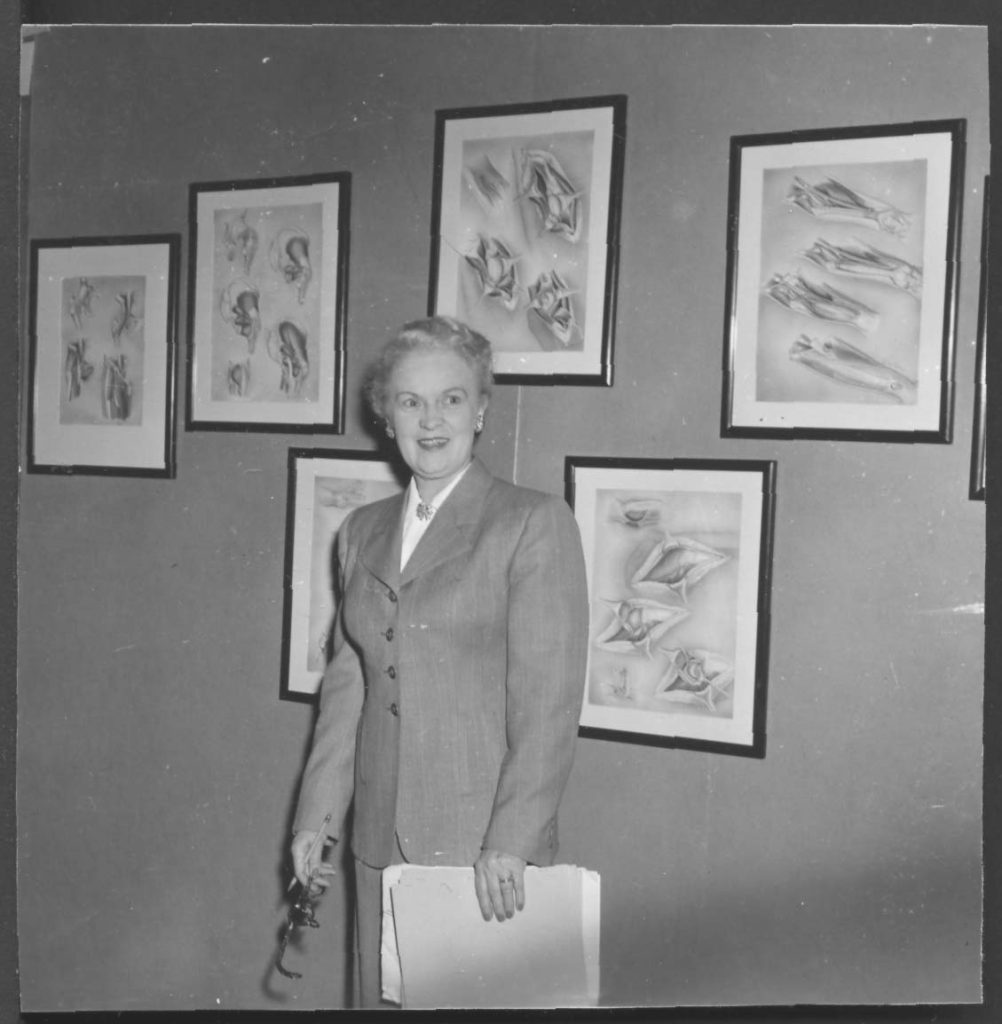
Winifred Rock in front of illustrations by Helen MacArthur, Surgery Department, Royal Victoria Hospital.
Advances in printing technology, especially the wider adoption of offset lithography in the 1940s and 1950s, made it possible to reproduce delicate tonal washes and full-colour plates at a reasonable cost. A closer look at her illustrated publications reveals the significance of her contributions at the Royal Victoria Hospital.

Gazette article reporting on the Textbook of Surgery, including MacArthur’s work.
The Textbook of Surgery, begun in 1944 and published in 1952, marks a landmark moment in Montreal’s medical history. Edited by Dr. H. F. Moseley, its first edition featured hundreds of MacArthur’s black and white illustrations as well as colour plates by Frank Netter.
Supported financially by Montreal philanthropist Miss Olive Hosmer, work on this book led to the formal establishment of the Department of Medical Illustration at RVH, granting increased status and security to illustrators who had been essential to teaching and research for decades. The textbook became a standard reference with continued revised editions.
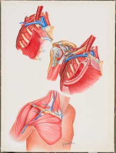
Illustration by Helen MacArthur for The Forequarter Amputation by Herbert Moseley.
A later collaboration with Moseley, Recurrent Dislocation of the Shoulder (1961), was the first publication of McGill University Press. MacArthur notably created illustrations in this volume in black and white as well as in colour.
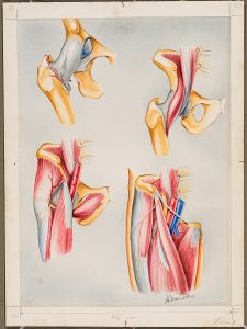
Illustration by Helen MacArthur for The Forequarter Amputation by Herbert Moseley.
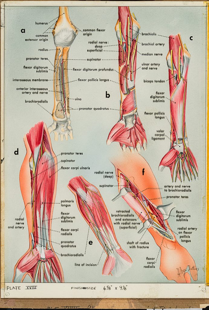
An illustration in colour could take up to two weeks, whereas working in black and white was much faster for MacArthur. Valuable as a tool for visual communication, she used heightened, contrasting colours to thoughtfully distinguish muscle layers, trace nerves, or highlight surgical fields against muted surrounding anatomy. Her priority was always accuracy, working from observation in the operating theatre and supplementing with a human skeleton kept in her studio and X-ray references.
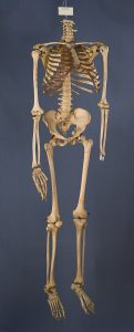
This Skeleton was used at the Department of Medical Illustration, Royal Victoria Hospital.
Press coverage of the Textbook of Surgery and her other works consistently credited MacArthur alongside authors and editors. Such acknowledgment, not commonly extended to medical illustrators, underscores that MacArthur’s work was regarded as equally vital as that of the authors.
This is likely in part due to Moseley’s advocacy, but MacArthur’s consistent signing of her illustrations suggests agency in her name appearing front and centre. MacArthur’s regular inclusion in publication credits no doubt helped foster greater respect and recognition for medical illustrators in Montreal and beyond.
The Fourquarter Amputation was published in 1957 and praised for its “wealth of illustrations.”