

The Art of Hortense Douglas: Cementing a place for Medical Illustration at the Montreal General Hospital
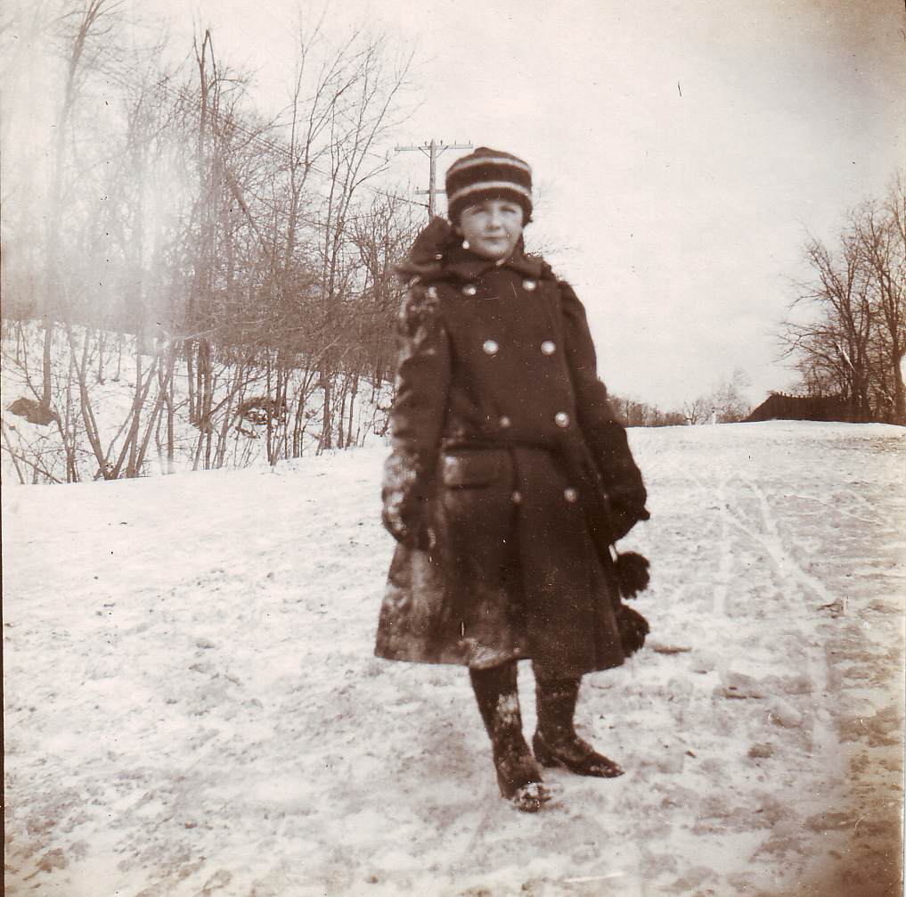
Hortense Douglas in a Red River coat, 1910
Hortense Douglas’ (1902-1979) professional arc as a medical illustrator crystallizes a transitional moment in Montreal medical history. Born in New York in 1901, her family soon moved to Montreal. Having become deaf as a child, she learned lip-reading and attended elite private school Miss Edgar and Miss Cramps. She studied charcoal drawing at the Montreal Art Association and later took life-drawing classes in New York.
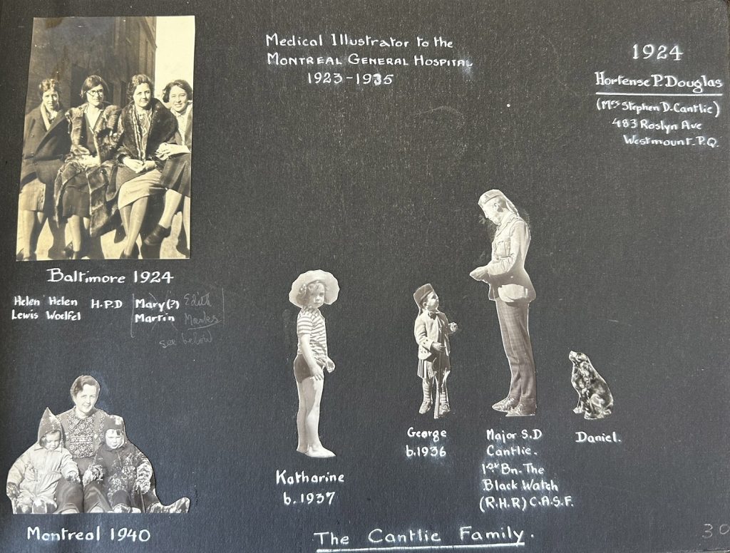
Biographic page by Hortense Douglas and her family from a scrapbook dedicated to Max Brödel upon his retirement.
As a Junior League member with fine arts training, she began volunteering at the Montreal General Hospital’s Pathology department in 1923, “making drawings of watercolours from specimens that were of special interest.” By 1925, she trained at Johns Hopkins Department of Arts Applied to medicine. She graduated and returned to her position in Montreal the following year as a formally trained and employed medical illustrator at the MGH.
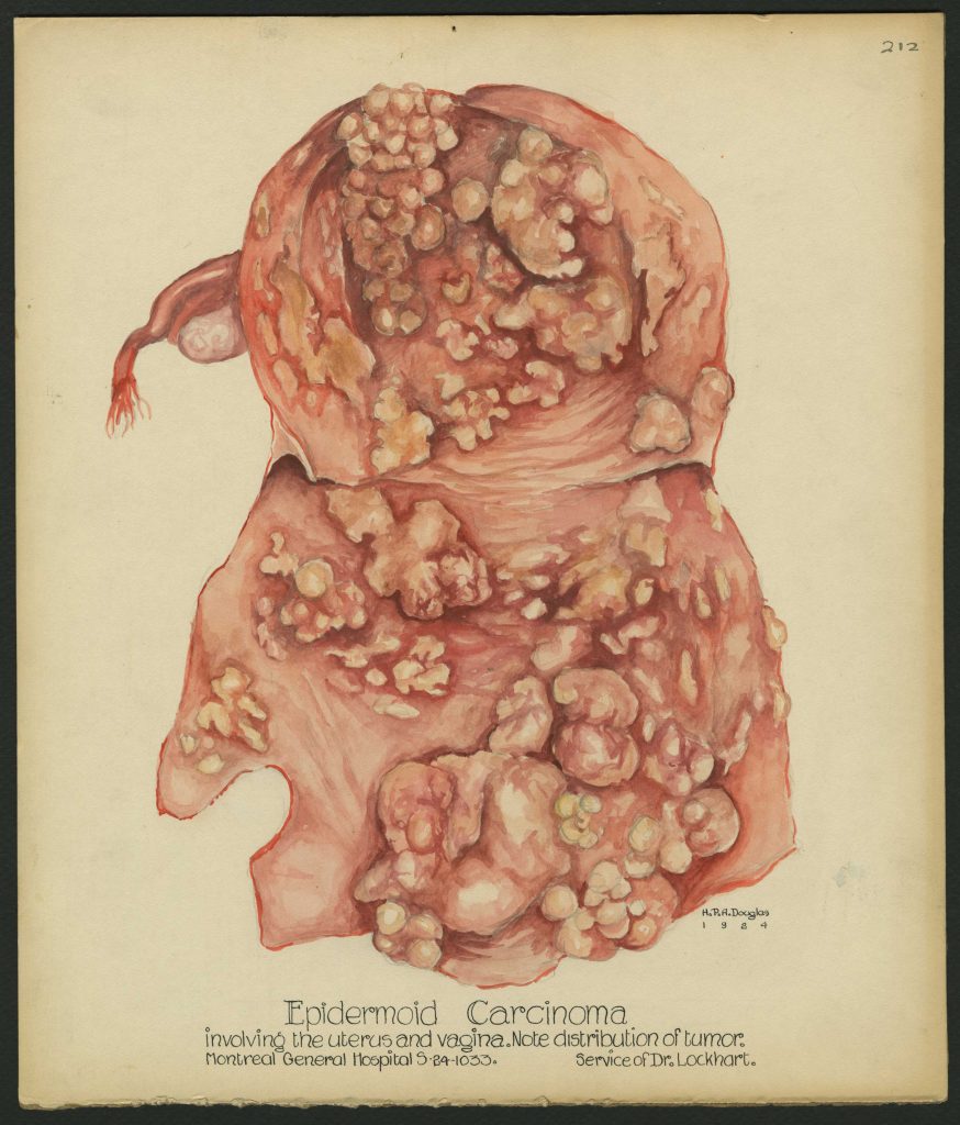
Illustration in watercolour by Hortense Douglas from 1924.
Illustration in watercolour by Hortense Douglas from 1924.
The hospital’s annual report from 1925 recognizes her work and underscores an important point:
A medical illustrator is not a luxury in any hospital; especially is this true of teaching hospitals.
The mid 1920s saw the “bench to bedside” model of medical education emerge. Science, research and practice were becoming equally essential parts of training. Douglas’ trajectory from volunteer to trained, staff illustrator at the MGH proves that medical illustration was gaining recognition in Montreal as essential to this new model.
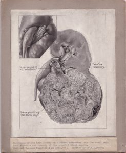
Illustration on stipple board by Hortense Douglas from 1926.
Douglas’ archives preserve examples before and after her training, as well as in-progress versions of work with neurologist Wilder Penfield. Her post-1925 drawings reveal her mastery of stippling and carbon dust—techniques that defined the field at the time. Even her drafting tools survive.
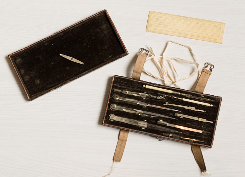
Drafting tools belonging to Hortense Douglas.
Her most reproduced work is the homunculus. This distorted human figure, mapped onto a visualization of a brain, illustrates how the brain’s cortex processes sensory information for different body parts. The image grew from Penfield’s brain mapping research of the 1930s–50s and became an icon of neuroscience.
Douglas, as Penfield’s principal illustrator, produced the first homunculus drawings for his books and articles. Her work was pivotal in visualizing the concept and cementing its widespread influence.
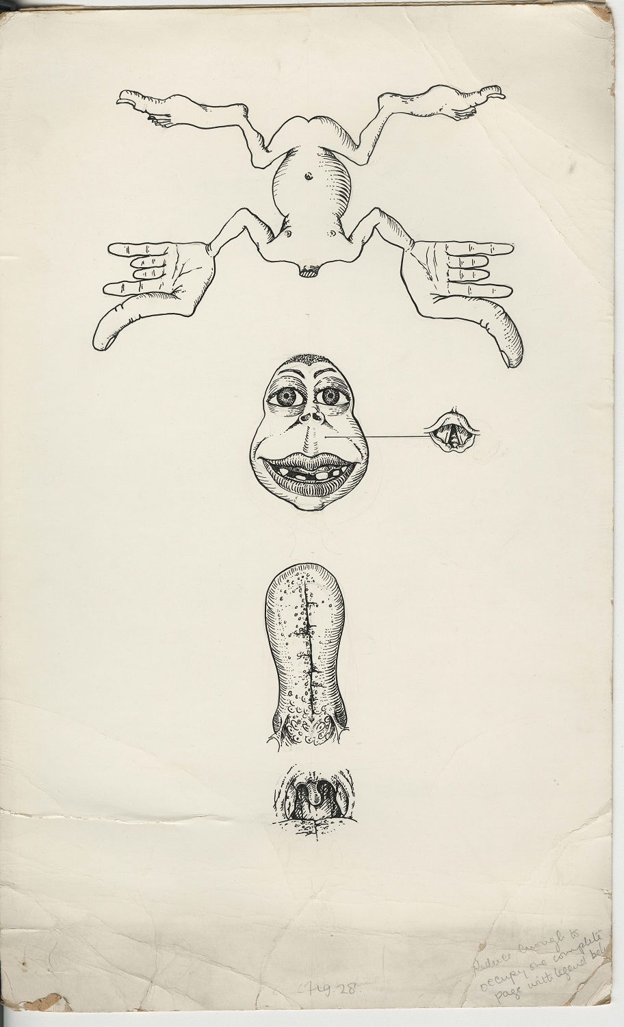
This is the first iteration of the homunculus that Hortense Douglas created while working with Penfield.